
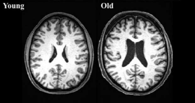
Brain scans from a 38-year-old, left, and a 73-year-old.
Researchers are embarking on a new study to answer how some people are able to stay sharper than others as they age.
Neurons in the brain that produce the pleasure-signaling neurotransmitter dopamine also directly control the brain’s circadian center, or “body clock” – the area that regulates eating cycles, metabolism and waking/resting cycles – a key link that possibly affects the body’s ability to adapt to jet lag and rotating shift work, a new study has demonstrated.
A molecule produced by insulating glial cells facilitates the functional wiring of brain cells involved in motor coordination.
Using the latest MRI scanning procedures, a team of researchers has shown how certain disorders of the hippocampus can initiate a drug resistant epilepsy. The team has discovered biomarkers that – if used for screening – could massively improve treatment options for epilepsy. The researchers have published their results in the online journal eLife.
A new study reveals the role circular RNA plays in brain function, including synaptic transmission and sensorimotor gating.
Depression has been shown to alter the structure of the brain’s white matter, which contains the circuitry that allows brain cells to communicate with each other, and which underpins brain function.
According to a new Nature study, in order for our taste system to work, the connection between neurons and taste bud cells have to rewire correctly each time.
A new optogenetic method called Optobow is helping researchers to discover specific and individual components of functional neural networks in the living brain. A Nature Communications report states this new method can help provide more detailed insights into both brain function and structure.
A neuroimaging study reveals people who report widespread pain have increased gray matter and functional connectivity in sensory and motor areas of the brain.
Finally this week, a large scale SPECT imaging study reveals women’s brains are significantly more active in more regions than males, including the prefronal cortex and limbic areas. Visual and areas associated with coordination were more active in males, researchers noted.
