Inside A Glass Brain

Philip Rosedale, creator of Second Life, and Adam Gazzaley, a neuroscientist at the University of California San Francisco, have developed a way for people to see each thought as it flies through the mind.  Entitled “The Glass Brain,” the project was on display at SXSW 2014 and provided attendees the opportunity to see how their brains react to assorted stimuli.

This is an anatomically-realistic 3D brain visualization depicting real-time source-localized activity (power and “effective” connectivity) from EEG (electroencephalographic) signals. Each color represents source power and connectivity in a different frequency band (theta, alpha, beta, gamma) and the golden lines are white matter anatomical fiber tracts. Estimated information transfer between brain regions is visualized as pulses of light flowing along the fiber tracts connecting the regions.

Weekly Neuroscience Update

For the study, researchers recruited twenty skilled pianists from Lyon, France. The group was asked to learn simple melodies by either hearing them several times or performing them several times on a piano. Pianists then heard all of the melodies they had learned, some of which contained wrong notes, while their brain electric signals were measured using electroencephalography (EEG). Credit: Caroline Palmer/Brian Mathias

For the study, researchers recruited twenty skilled pianists from Lyon, France. The group was asked to learn simple melodies by either hearing them several times or performing them several times on a piano. Pianists then heard all of the melodies they had learned, some of which contained wrong notes, while their brain electric signals were measured using electroencephalography (EEG). Credit: Caroline Palmer/Brian Mathias

Research from McGill University reveals that the brain’s motor network helps people remember and recognize music that they have performed in the past better than music they have only heard. 

Veterans who have been exposed to nearby explosions, but who lack clear symptoms of traumatic brain injury (TBI) may still have damage to the brain’s white matter similar to veterans with TBI, according to new research at Duke Medicine and the U.S. Department of Veterans Affairs.

University of Adelaide researchers say new insights into how the human brain responds to chronic pain could eventually lead to improved treatments for patients. And also at the University of Adelaide, studies show that a gene linked to intellectual disability is critical to the earliest stages of the development of human brains. Known as USP9X, the gene has been investigated by Adelaide researchers for more than a decade, but in recent years scientists have begun to understand its particular importance to brain development.

In order to function properly, the human brain requires the ability not only to store but also to forget. Through memory loss, unnecessary information is deleted and the nervous system retains its plasticity. A disruption of this process can lead to serious mental disorders. Scientists have now discovered a molecular mechanism that actively regulates the process of forgetting. The journal Cell has published their results.

Brain imaging using radioactive dye can detect early evidence of Alzheimer’s disease that may predict future cognitive decline.

Scientists have identified a new group of compounds that may protect brain cells from inflammation linked to seizures and neurodegenerative diseases.

Your brain’s ability to instantly link what you see with what you do is down to a dedicated information ‘highway’, suggests new research.

There has been a great deal of research on changes among different brain states during sleep, but  new findings, reported in the March 13 issue of Cell, provide a compelling example of a change in state in the awake brain.

Finally, this week, using a new method to determine whether individuals met the criteria for a clinical diagnosis of bipolar disorder, researchers from UCLA tried a new approach by combining results from brain imaging, cognitive tests, and an array of temperament and behavioral measures. In an attempt to better understand the genes that cause the disorder, a collaborative research team identified about 50 brain and behavioral measures that are both under strong genetic control and associated with bipolar disorder, creating the potential to pinpoint the specific genes that contribute to the illness.

Inside The Decision-Making Brain

anterior_ insula

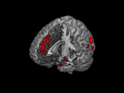
The anterior insula was sensitive to escalating alcohol costs especially when the costs of drinking outweighed the benefits, indicating this could be the region of the brain at the intersection of how our rational and irrational systems work with one another.

Although choosing to do something because the perceived benefit outweighs the financial cost is something people do daily, little is known about what happens in the brain when a person makes these kinds of decisions. Studying how these cost-benefit decisions are made when choosing to consume alcohol, University of Georgia associate professor of psychology James MacKillop identified distinct profiles of brain activity that are present when making these decisions.*

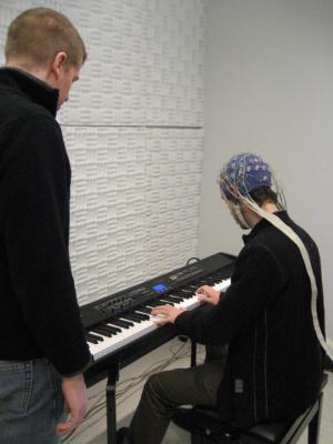
The study combined functional magnetic resonance imaging and a bar laboratory alcohol procedure to see how the cost of alcohol affected people’s preferences. The study group included 24 men, age 21-31, who were heavy drinkers. Participants were given a $15 bar tab and then were asked to make decisions in the fMRI scanner about how many drinks they would choose at varying prices, from very low to very high. Their choices translated into real drinks, at most eight that they received in the bar immediately after the scan. Any money not spent on drinks was theirs to keep.

The study applied a neuroeconomic approach, which integrates concepts and methods from psychology, economics and cognitive neuroscience to understand how the brain makes decisions. In this study, participants’ cost-benefit decisions were categorized into those in which drinking was perceived to have all benefit and no cost, to have both benefits and costs, and to have all costs and no benefits. In doing so, MacKillop could dissect the neural mechanisms responsible for different types of cost-benefit decision-making.

When participants decided to drink in general, activation was seen in several areas of the cerebral cortex, such as the prefrontal and parietal cortices. However, when the decision to drink was affected by the cost of alcohol, activation involved frontostriatal regions, which are important for the interplay between deliberation and reward value, suggesting suppression resulting from greater cognitive load. This is the first study of its kind to examine cost-benefit decision-making for alcohol and was the first to apply a framework from economics, called demand curve analysis, to understanding cost-benefit decision making.

The brain activity was most differentially active during the suppressed consumption choices, suggesting that participants were experiencing the most conflict. We had speculated during the design of the study that the choices not to drink at all might require the most cognitive effort, but that didn’t seem to be the case. Once people decided that the cost of drinking was too high, they didn’t appear to experience a great deal of conflict in terms of the associated brain activity. McKillop

These conflicted decisions appeared to be represented by activity in the anterior insula, which has been linked in previous addiction studies to the motivational circuitry of the brain. Not only encoding how much people crave or value drugs, this portion of the brain is believed to be responsible for processing interceptive experiences, a person’s visceral physiological responses.

It was interesting that the insula was sensitive to escalating alcohol costs especially when the costs of drinking outweighed the benefits. That means this could be the region of the brain at the intersection of how our rational and irrational systems work with one another. In general, we saw the choices associated with differential brain activity were those choices in the middle, where people were making choices that reflect the ambivalence between cost and benefits. Where we saw that tension, we saw the most brain activity. McKillop

While MacKillop acknowledges the impact this research could have on neuromarketing–or understanding how the brain makes decisions about what to buy–he is more interested in how this research can help people with alcohol addictions.

“These findings reveal the distinct neural signatures associated with different kinds of consumption preferences. Now that we have established a way of studying these choices, we can apply this approach to better understanding substance use disorders and improving treatment,” he said, adding that comparing fMRI scans from alcoholics with those of people with normal drinking habits could potentially tease out brain patterns that show what is different between healthy and unhealthy drinkers. “In the past, we have found that behavioral indices of alcohol value predict poor treatment prognosis, but this would permit us to understand the neural basis for negative outcomes.”

*The research was published in the journal Neuropsychopharmacology March 3.

Weekly Neuroscience Update

Embed from Getty Images

Johns Hopkins researchers report that people with chronic insomnia show more plasticity and activity than good sleepers in the part of the brain that controls movement.

Researchers at The Scripps Research Institute (TSRI) and Vanderbilt University have created the most detailed 3-D picture yet of a membrane protein that is linked to learning, memory, anxiety, pain and brain disorders such as schizophrenia, Parkinson’s, Alzheimer’s and autism.

New research has revealed the dramatic effect the immune system has on the brain development of young children. The findings suggest new and better ways to prevent developmental impairment in children in developing countries, helping to free them from a cycle of poverty and disease, and to attain their full potential.

Rate of change in the thickness of the brain’s cortex is an important factor associated with a person’s change in IQ, according to a collaborative study by scientists in five countries.

Researchers have found that decision-making accuracy can be improved by postponing the onset of a decision by a mere fraction of a second. The results could further our understanding of neuropsychiatric conditions characterized by abnormalities in cognitive function and lead to new training strategies to improve decision-making in high-stake environments. The study was published in the March 5 online issue of the journal PLoS One.

A study has revealed how the fatal neurodegenerative disease amyotrophic lateral sclerosis (ALS), also known as Lou Gehrig’s disease, is transmitted from cell to cell, and suggests the spread of the disease could be blocked.

Research from Karolinska Institutet in Sweden suggests that the expression of the so called MYC gene is important and necessary for neurogenesis in the spinal cord. The findings are being published in the journal  EMBO Reports .

Our memories are inaccurate, more than we’d like to believe. And now a study demonstrates one reason: we apparently add current experiences onto memories.

Damage to the brain may still occur even if symptoms of traumatic brain injury are not present, scientists suggest.

The brain processes read and heard language differently. This is the key and new finding of a study published in Frontiers in Human Neuroscience.

How Your Brain Sees Things You Don’t

What do you see in this image?  (Credit: Jay Sanguinetti)

What do you see in this image? (Credit: Jay Sanguinetti)

A new study indicates that our brains perceive objects in everyday life that we may not be consciously aware of.

The finding by University of Arizona doctoral student Jay Sanguinetti challenges currently accepted models, in place for a century, about how the brain processes visual information.

Sanguinetti showed study participants a series of black silhouettes, some of which contained meaningful, real-world objects hidden in the white spaces on the outsides. He monitored subjects’ brainwaves with an electroencephalogram, or EEG, while they viewed the objects.

Study participants’ brainwaves indicated that even if a person never consciously recognized the shapes on the outside of the image, their brains still processed those shapes to the level of understanding their meaning.

N400-reduction

A brainwave that indicates recognition of an object

“There’s a brain signature for meaningful processing,” Sanguinetti said. A peak in the averaged brainwaves called N400 indicates that the brain has recognized an object and associated it with a particular meaning.

“It happens about 400 milliseconds after the image is shown, less than a half a second,” said Peterson. “As one looks at brainwaves, they’re undulating above a baseline axis and below that axis.

The negative ones below the axis are called N and positive ones above the axis are called P, so N400 means it’s a negative waveform that happens approximately 400 milliseconds after the image is shown.”

The presence of the N400 negative peak indicates that subjects’ brains recognize the meaning of the shapes on the outside of the figure.

“The participants in our experiments [in some cases] don’t see those shapes on the outside; nonetheless, the brain signature tells us that they have processed the meaning of those shapes,” said said Sanguinetti adviser Mary Peterson, a professor of psychology and director of the UA’s Cognitive Science Program.

“But the brain rejects them as interpretations, and if it rejects the shapes from conscious perception, then you won’t have any awareness of them.”

“We also have novel silhouettes as experimental controls,” Sanguinetti said. “These are novel black shapes in the middle and nothing meaningful on the outside.”

The N400 waveform does not appear on the EEG of subjects when they are seeing these truly novel silhouettes, without images of any real-world objects, indicating that the brain does not recognize a meaningful object in the image.

“This is huge,” Peterson said. “We have neural evidence that the brain is processing the shape and its meaning of the hidden images in the silhouettes we showed to participants in our study.”

So why does the brain process images that are not perceived?

The finding leads to the question: why would the brain process the meaning of a shape when a person is ultimately not going to perceive it?

“Many, many theorists assume that because it takes a lot of energy for brain processing, that the brain is only going to spend time processing what you’re ultimately going to perceive,” said Peterson.

“But in fact the brain is deciding what you’re going to perceive, and it’s processing all of the information and then it’s determining what’s the best interpretation.

“This is a window into what the brain is doing all the time. It’s always sifting through a variety of possibilities and finding the best interpretation for what’s out there. And the best interpretation may vary with the situation.”

Our brains may have evolved to sift through the barrage of visual input in our eyes and identify those things that are most important for us to consciously perceive, such as a threat or resources such as food, Peterson suggested.

Finding where the processing of meaning occurs

In the future, Peterson and Sanguinetti plan to look for the specific regions in the brain where the processing of meaning occurs to understand where and how this meaning is processed,” said Peterson.

Images were shown to Sanguinetti’s study participants for only 170 milliseconds, yet their brains were able to complete the complex processes necessary to interpret the meaning of the hidden objects.

“There are a lot of processes that happen in the brain to help us interpret all the complexity that hits our eyeballs,” Sanguinetti said. “The brain is able to process and interpret this information very quickly.”

How this relates to the real world

Sanguinetti’s study indicates that in our everyday life, as we walk down the street, for example, our brains may recognize many meaningful objects in the visual scene, but ultimately we are aware of only a handful of those objects, said Sanguinetti.

The brain is working to provide us with the best, most useful possible interpretation of the visual world — an interpretation that does not necessarily include all the information in the visual input.

“The findings in the research also show that our brains are processing potential objects in a visual scene to much higher levels of processing than once thought,” he explained to KurzweilAI. “Our models assume that potential objects compete for visual representation. The one that wins the competition is perceived as the object, the loser is perceived as the shapeless background.

“Since we’ve shown that shapeless backgrounds are processed to the level of semantics (meaning), there might be a way to bias this processing such that hidden objects in a scene might be perceived, by tweaking the image in ways to enunciate certain objects over others. This could be useful in many applications like radiology, product design, and even art.”

Notes:

Silhouette Image: Sanguinetti showed study participants images of what appeared to be an abstract black object. Sometimes, however, there were real-world objects hidden at the borders of the black silhouette. In this image, the outlines of two seahorses can be seen in the white spaces surrounding the black object.

Original source of article  http://www.kurzweilai.net/does-your-brain-see-things-you-dont

REFERENCES:

Joseph L. Sanguinetti, John J. B. Allen, and Mary A. Peterson, The Ground Side of an Object: Perceived as Shapeless yet Processed for Semantics, Psychological Science, 2013, doi: 10.1177/0956797613502814

Weekly Neuroscience Update

Lesion overlap map illustrating common and distinctive brain regions for Val/Val (blue) and Val/Met (yellow) genotype patients. Overlap between Val/Val and Val/Met genotype patients is illustrated in green. In each axial slice, the right hemisphere is on the reader’s left. Credit Barbey et al./PLOS ONE.

Lesion overlap map illustrating common and distinctive brain regions for Val/Val (blue) and Val/Met (yellow) genotype patients. Overlap between Val/Val and Val/Met genotype patients is illustrated in green. In each axial slice, the right hemisphere is on the reader’s left – this is because the view is from the bottom i.e. as if one were in the neck looking up at the brain. Credit Barbey et al./PLOS ONE.

Researchers report that one tiny variation in the sequence of a gene may cause some people to be more impaired by traumatic brain injury (TBI) than others with comparable wounds.

A recent study conducted by a multicenter-research team led by Cedars-Sinai Medical Center used a new, automated imaging system to identify shrinkage of a mood-regulating brain structure in a large sample of women with multiple sclerosis (MS), who also have a certain type of depression. The research supports earlier studies suggesting that the hippocampus may contribute to the high frequency of depression seen in those who suffer from MS.

Max Ortiz Catalan, researcher at Chalmers University of Technology, has developed a new method for the treatment of phantom limb pain (PLP) after an amputation. 

University of Sydney study is looking into the effectiveness of omega-3 supplements and the antidepressant, sertraline, in reducing depressive symptoms and cognitive decline in older people, in a bid to prevent the onset of depression and dementia in later life.

Certain neurons in the human striatum—a brain region involved in movement and cognition—are renewed throughout life, according to a study published in Cell.

Researchers at the University of Oxford have discovered the molecular switch in the brain that sends us to sleep.

Can the damaged brain repair itself?

After a traumatic brain injury, it sometimes happens that the brain can repair itself, building new brain cells to replace damaged ones. But the repair doesn’t happen quickly enough to allow recovery from degenerative conditions like motor neuron disease (also known as Lou Gehrig’s disease or ALS). In this video, regenerative neurologist  Siddharthan Chandran walks through some new techniques using special stem cells that could allow the damaged brain to rebuild faster.


What Songbirds Can Teach Us About Learning and the Brain

All known languages require the following features: 

1.  Sufficient brain space to house the dictionary and grammar.

2. Specific features of the vocal apparatus including the vocal cords, the muscles of the tongue and mouth enabling articulation.

3. An ability to control breathing which allows for long fluent articulate phrases and the ability to modulate intonation subtly over the length of a single breadth.

Our nearest primate relatives (i.e. monkeys and apes) do not have any such control which explains why attempts to train them to speak have been so unsuccessful.

Birds alone can imitate human speech. The birds brain, vocal apparatus, or syrinx (literally, ‘flute’) and their ability to control their breathing explains why.


Weekly Neuroscience Update

Temporo-parietal jonction (TPJ) © Perrine Ruby / Inserm

Temporo-parietal jonction (TPJ) © Perrine Ruby / Inserm

Some people recall a dream every morning, whereas others rarely recall one. A research team has studied the brain activity of these two types of dreamers in order to understand the differences between them. In a study published in the journal Neuropsychopharmacology, the researchers show that the temporo-parietal junction, an information-processing hub in the brain, is more active in high dream recallers. Increased activity in this brain region might facilitate attention orienting toward external stimuli and promote intrasleep wakefulness, thereby facilitating the encoding of dreams in memory.

Many psychiatric disorders are accompanied by memory deficits. Basel scientists have now identified a network of genes that controls fundamental properties of neurons and is important for human brain activity, memory and the development of schizophrenia. 

Researchers have taken a major step toward identifying the specific genes that contribute to bipolar disorder.

A recent study conducted by Johns Hopkins University and the National Institute of Aging found that aging adults with hearing loss are at higher risk for accelerated brain-tissue loss.

Brain cell regeneration has been discovered in a new location in human brains. The finding raises hopes that these cells could be used to help people recover after a stroke, or to treat other brain diseases.

Finally this week, researchers are hoping that the world’s largest simulated brain — known as Spaun — will be used to test new drugs that lead to breakthrough treatments for neurological disorders such as Parkinson’s, Huntington’s, and Alzheimer’s disease.