Weekly Neuroscience Update

Temporo-parietal jonction (TPJ) © Perrine Ruby / Inserm

Temporo-parietal jonction (TPJ) © Perrine Ruby / Inserm

Some people recall a dream every morning, whereas others rarely recall one. A research team has studied the brain activity of these two types of dreamers in order to understand the differences between them. In a study published in the journal Neuropsychopharmacology, the researchers show that the temporo-parietal junction, an information-processing hub in the brain, is more active in high dream recallers. Increased activity in this brain region might facilitate attention orienting toward external stimuli and promote intrasleep wakefulness, thereby facilitating the encoding of dreams in memory.

Many psychiatric disorders are accompanied by memory deficits. Basel scientists have now identified a network of genes that controls fundamental properties of neurons and is important for human brain activity, memory and the development of schizophrenia. 

Researchers have taken a major step toward identifying the specific genes that contribute to bipolar disorder.

A recent study conducted by Johns Hopkins University and the National Institute of Aging found that aging adults with hearing loss are at higher risk for accelerated brain-tissue loss.

Brain cell regeneration has been discovered in a new location in human brains. The finding raises hopes that these cells could be used to help people recover after a stroke, or to treat other brain diseases.

Finally this week, researchers are hoping that the world’s largest simulated brain — known as Spaun — will be used to test new drugs that lead to breakthrough treatments for neurological disorders such as Parkinson’s, Huntington’s, and Alzheimer’s disease.
Why Are Some Brain Injuries Worse Than Others?

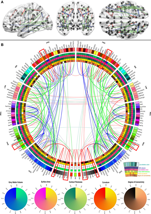
Image A: 3-D models of how the white matter in the brain connects, paired with a "connectogram" visualizing linkages between different areas of the brain / USC

3-D models of how the white matter in the brain connects, paired with a “connectogram” visualizing linkages between different areas of the brain / USC

According to research published in The Lancet, approximately a fifth of adults with a severe traumatic brain injury make a good recovery. But many more die or are left with enduring disability. Although doctors caring for Michael Schumacher, the Formula One World Champion who sustained a severe head injury while skiing, haven’t commented on how he is responding to their latest tests and treatment, Dr Peter Kirkpatrick, a leading British neurosurgeon based at Addenbrooke’s hospital in Cambridge, says that it is “extremely unlikely” that Schumacher will return to his previous level of health, although he insists it is “medically possible”.

The effects of brain injury fall into three main categories:

  • Cognitive – problems with memory, concentration, information processing
  • Emotional and behavioural problems – anxiety, explosive anger and irritability, lack of awareness or empathy
  • Physical – problems with movement, balance and co-ordination, fatigue, epilepsy

Sometimes a head injury which seems severe is followed by a good recovery while a seemingly small head injury can have very serious, long-lasting consequences.  Why is this?

Location, location, location.

The reason is that brain injury operates a bit like the property market in that the three most important things to consider are location, location and location. When nerve pathways are damaged, those brain areas served by those pathways may wither or have their functions taken over by other brain regions. Nerve pathways are also called ‘white’ pathways or ‘white matter’ because they are covered by an insulating sheath of myelin and appear white to the naked eye.

The challenge is to determine the location of key ‘scaffold’ pathways and to understand what makes them so vulnerable and important. This is not an easy task given the total length of nerve pathways in the average 20-year old human brain is 160,000 km. A recent study provides new findings on the brain’s network scaffold that will help inform clinicians about the neurological impacts of brain diseases such as multiple sclerosis, Alzheimer’s disease and brain injury.


Weekly Neuroscience Update

brain

Researchers have pinpointed an area of the brain that serves as an arbitrator between our two decision-making systems, one devoted to habitual behaviors and the other devoted to actions we take only after careful consideration.

Scientists who discovered a gene that links the thickness of the brain’s gray matter to intelligence say their finding might help improve understanding of brain disorders such as autism and schizophrenia.

People who appreciate the beauty of mathematics activate the same part of their brain when they look at aesthetically pleasing formula as others do when appreciating art or music, suggesting that there is a neurobiological basis to beauty.

Researchers at the University of California, San Francisco, are looking at ways video game technology can be used to improve or heal the brain.

For the first time, scientists have identified a gene linking the thickness of the grey matter in the brain to intelligence. The study is published today in Molecular Psychiatry and may help scientists understand biological mechanisms behind some forms of intellectual impairment.

People with dyslexia have greater difficulty than others in shifting attention between the senses – from seeing something to hearing something – according to an Oxford University study. The researchers suggest that action video games, where attention is constantly shifting focus, may be able to boost literacy skills – though they have yet to test this. The findings are reported in the journal Current Biology.

A new study published in Social Neuroscience says that the brain has begun processing a typed emoticon with the same signals that previously only accompanied processing a real, emoting human face.

A specific type of neuron in the amygdala performs differently in individuals with autism spectrum disorder than in those without the disorder, according to a new study by researchers at Cedars-Sinai Medical Center in New York.

Finally, this Valentine’s weekend, new research reveals that  a region deep inside the brain controls how quickly people make decisions about love.

Does the brain rewrite memories?

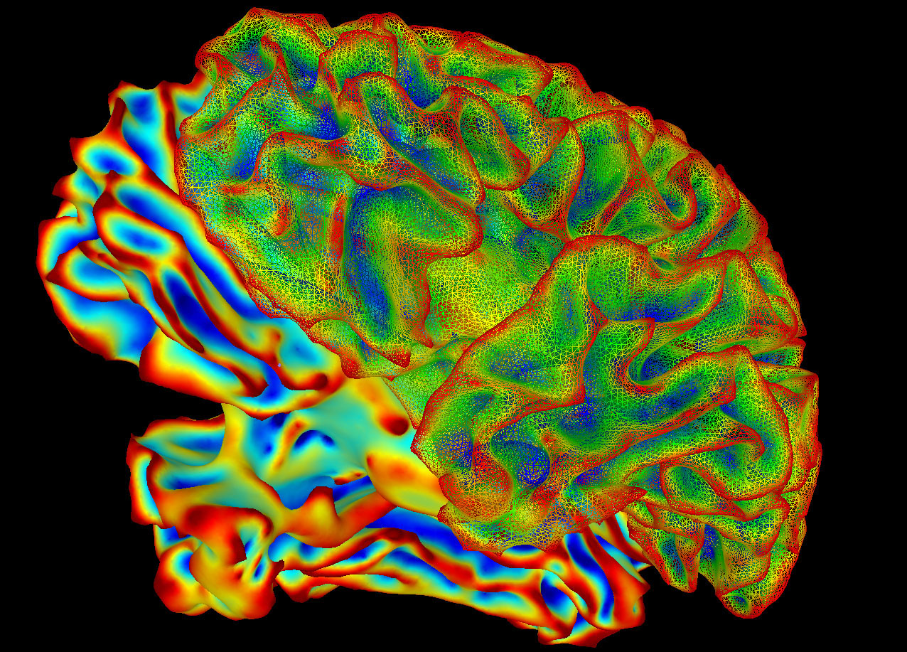
A multi-colored image of a brain for imaging research. Researchers from Northwestern University released a study showing that the memories can adapt. Scientists used a functional MRI and eye tracking as part of their research.

A multi-colored image of a brain for imaging research. Researchers from Northwestern University released a study showing that the memories can adapt. Scientists used a functional MRI and eye tracking as part of their research.

Memories are not picture perfect. The brain can rewrite old memories to update them with new experiences and information, according to a new study by Northwestern University scientists. The study, published in the Journal of Neuroscience, monitored 17 participants using three different tools: behavioral measures, a functional MRI and eye-movement tracking. Despite the number of participants appearing small, the researchers said it is a statistically robust study.

Read the full story here 

The Influence of Taking Photos on Memory

medium_4786580516

Taking a picture to help you remember something might end up having the opposite effect, according to new research.

A study showed that people who took photographs of items during a museum tour were less likely to remember details than those who merely looked at the objects.

“People so often whip out their cameras almost mindlessly to capture a moment, to the point that they are missing what is happening right in front of them,” says psychological scientist Linda Henkel, author of the study, which was published in the journal Psychological Science.

Henkel set up an experiment in the university’s museum, in which students were led on a tour and were asked to take note of certain objects, either by photographing them or by simply observing them. The next day, their memory for the objects was tested – and participants were less accurate in recognising the items they had photographed compared to those they had only observed. Henkel called this the “photo-taking impairment effect.”

“When people rely on technology to remember for them – counting on the camera to record the event and thus not needing to attend to it fully themselves – it can have a negative impact on how well they remember their experiences,” she says.

A second group offered a slight variation on the findings: those taking a photograph of a specific detail on the object by zooming in on it with the camera seemed to preserve memory for the object, not just for the part that was zoomed in on but also for the part that was out of frame.

“These results show how the ‘mind’s eye’ and the camera’s eye are not the same,” Henkel says, adding that memory research indicates taking pictures can help people remember, but only if they take time to observe and review.

photo credit: Carollainy via photopin cc

Weekly Neuroscience Update

The salience network, highlighted here in two epilepsy patients, is thought to mediate our response to important internal or external signals, such as pain or the sound of a siren. Image: Parvizi et al. Neuron 2013

The salience network, highlighted here in two epilepsy patients, is thought to mediate our response to important internal or external signals, such as pain or the sound of a siren. Image: Parvizi et al. Neuron 2013

In a rare study involving direct brain stimulation researchers say they have uncovered direct evidence that a brain region known as the anterior midcingulate cortex and its surrounding network play a central role in motivation and a readiness to act.

Many studies suggest that pushing your brain to multitask—writing emails, for instance, while watching the day’s latest news and eating breakfast—leads to poorer performance and lower productivity. But for at least one everyday task—visual sampling (the act of picking up bits of visual information through short glances)—multitasking is not a problem for the brain. A collaboration between researchers at the UC Santa Barbara and the University of Bristol in the UK has shown that during visual sampling, the brain can handle various visual functions simultaneously.

Researchers report a detailed account of how speech sounds are identified by the human brain, offering an unprecedented insight into the basis of human language. The finding, they said, may add to our understanding of language disorders, including dyslexia.

A new study correlating brain activity with how people make decisions suggests that when individuals engage in risky behavior, it’s probably not because their brains’ desire systems are too active, but because their self-control systems are not active enough. This might have implications for how health experts treat mental illness and addiction or how the legal system assesses a criminal’s likelihood of committing another crime.

Pain sensitivity is controlled by a genetic “dimmer switch”, which can be re-set, UK scientists have discovered.

Amputee Feels in Real-Time with Bionic Hand

Dennis Aabo Sørensen is the first amputee in the world to feel sensory rich information — in real-time — with a prosthetic hand wired to nerves in his upper arm. Sørensen could grasp objects intuitively and identify what he was touching while blindfolded.
Sørensen participated in the clinical study for one month in early 2013. The results are published in Science Translational Medecine.

Read more on this:
http://bit.ly/201402EPFL_BionicHand
http://bit.ly/2014EPFLYoutube_BionicHand
http://stm.sciencemag.org/

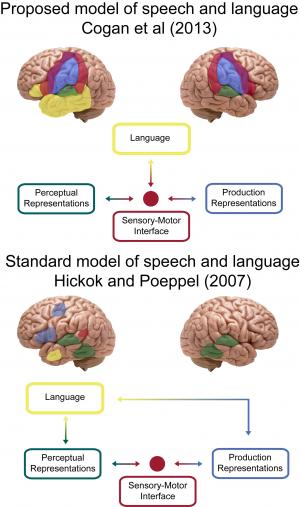
Speech processing requires both sides of our brain

A new study by Cogan et al proposes that speech processes occur on both sides of the brain and are distinct from language, which occurs on one side, typically on the left. This suggests a revision to the standard model of how speech is linked to language with some processes going through a "bilateral sensory-motor interface". Credit: Greg Cogan and Bijan Pesaran

A new study by Cogan et al proposes that speech processes occur on both sides of the brain and are distinct from language, which occurs on one side, typically on the left. This suggests a revision to the standard model of how speech is linked to language with some processes going through a “bilateral sensory-motor interface”. Credit: Greg Cogan and Bijan Pesaran

A new study has found that we use both sides of our brain for speech, a finding that alters previous conceptions about neurological activity. The results, which appear in the journal Nature, also offer insights into addressing speech-related inhibitions caused by stroke or injury and lay the groundwork for better rehabilitation methods.

The study’s senior author, Bijan Pesaran, an associate professor in New York University’s Center for Neural Science, said:

Our findings upend what has been universally accepted in the scientific community—that we use only one side of our brains for speech. With this greater understanding of the speech process, we can retool rehabilitation methods in ways that isolate speech recovery and that don’t involve language.

Read more: Sensory–motor transformations for speech occur bilaterally