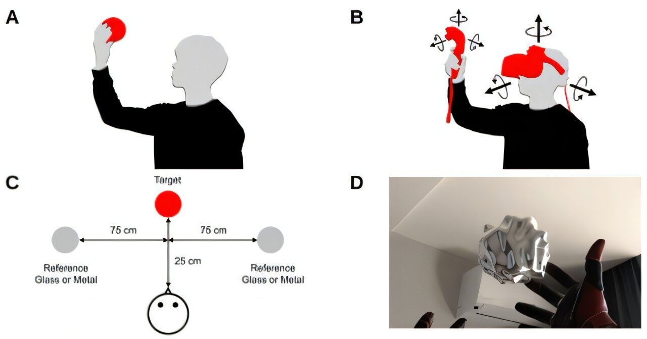
Researchers have conducted a psychophysical study using virtual reality (VR) to investigate how humans flexibly use exploratory behaviours—such as changing their viewpoint by moving their head and manipulating objects with their hands—when discriminating the material properties of objects. The study was published in the Journal of Vision.
A new study establishes a robust link between long-term exposure to air pollution and the risk of Parkinson’s disease.
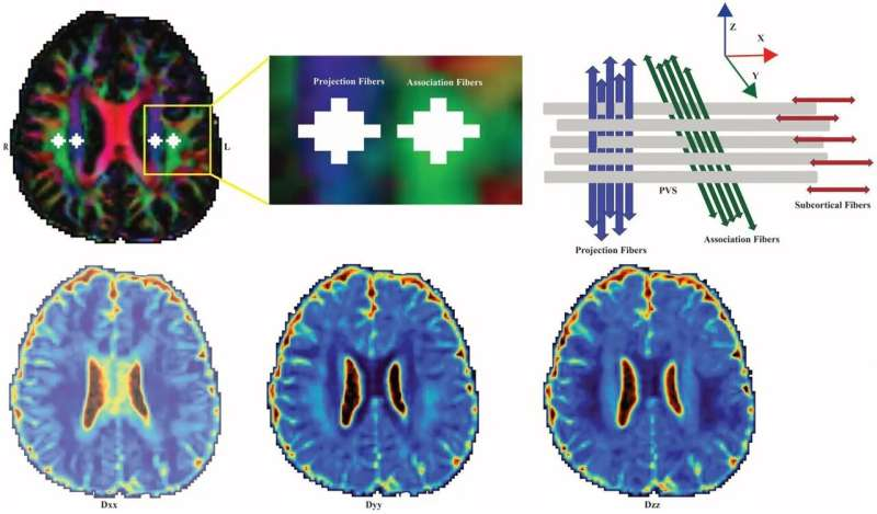
An international team of scientists and clinicians have developed a generative artificial intelligence framework that unmasks these previously hidden cortical lesions by analysing existing legacy MRI scans. By synthesising minor, sub-visual discrepancies across multiple image contrasts, the AI acts as a computational lens, extracting vital diagnostic data from ordinary scans and revealing an entirely invisible layer of MS pathology.
New research in General Psychiatry has uncovered a link between higher levels of daytime light exposure and a lower risk of dementia.
A recent study discovered that randomly played sounds during sleep can actively impair memory consolidation. By utilising real-time electroencephalography (EEG) monitoring, the researchers proved that ambient sound clicks disrupt deep slow-wave sleep, arresting the physical propagation of slow brain waves across the cortex and fracturing the crucial transfer of information required to form long-term memories.
A research team has uncovered new insight into how the brain senses movement. Their findings could potentially help improve sensation and movement for prosthetic limbs.
A new study has demonstrated that introducing inflammatory signalling molecules directly into human hippocampal stem cells halts new neuron production. Instead of simply dying or becoming damaged, the brain’s neural stem cells abandon their regenerative responsibilities, entering an “immune alert” state that fuels localised neuroinflammation.
People who speak more than one language seem to have younger brains, according to research presented at the Federation of European Neuroscience Societies (FENS) Forum 2026.
A new research framework that combines large language models (LLMs) with choice mathematics to evaluate human decision-making. By deploying LLMs to automatically interpret and code thousands of free-text participant thought justifications, the framework provides a scalable, validated methodology demonstrating that human reasoning strategies shift dynamically with a problem’s structure.
Scientists have found that human cortical neurons function like advanced microchips, demonstrating computational abilities comparable to those of deep artificial neural networks, rather than merely acting as simple switches.