Australian cricketer, Phillip Hughes.
Australian cricketer Phillip Hughes has died two days after being struck in the head by a bouncer while batting for South Australia at the The Sydney Cricket Ground. Hughes, 25, had been in an induced coma since the accident on Tuesday afternoon.
Two of the most tragic events within Australian cricket in just over the past decade have involved catastrophic head injuries: firstly to David Hookes in a hotel altercation, secondly to Phillip Hughes while batting. According to research published in The Lancet, approximately a fifth of adults with a severe traumatic brain injury make a good recovery. But many more die or are left with enduring disability.
So why are some brain injuries worse than others?
The effects of brain injury fall into three main categories:
- Cognitive – problems with memory, concentration, information processing
- Emotional and behavioural problems – anxiety, explosive anger and irritability, lack of awareness or empathy
- Physical – problems with movement, balance and co-ordination, fatigue, epilepsy
Sometimes a head injury which seems severe is followed by a good recovery while a seemingly small head injury can have very serious, long-lasting consequences. Why is this?
Location, location, location.
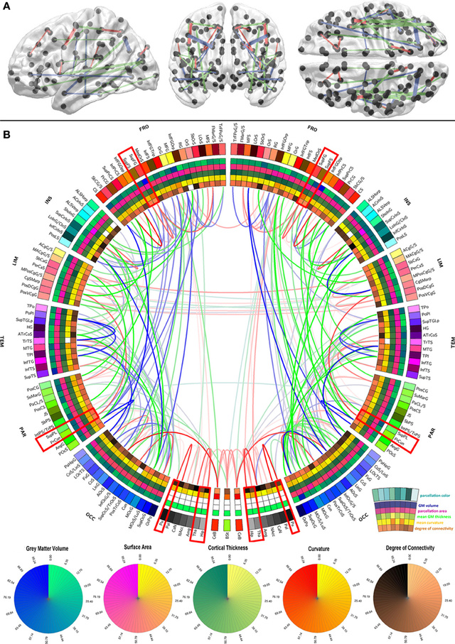
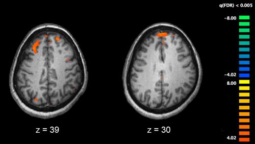
The reason is that brain injury operates a bit like the property market in that the three most important things to consider are location, location and location. When nerve pathways are damaged, those brain areas served by those pathways may wither or have their functions taken over by other brain regions. Nerve pathways are also called ‘white’ pathways or ‘white matter’ because they are covered by an insulating sheath of myelin and appear white to the naked eye.
The challenge is to determine the location of key ‘scaffold’ pathways and to understand what makes them so vulnerable and important. This is not an easy task given the total length of nerve pathways in the average 20-year old human brain is 160,000 km. A recent study provides new findings on the brain’s network scaffold that will help inform clinicians about the neurological impacts of brain diseases such as multiple sclerosis, Alzheimer’s disease and brain injury.