Three experiments to test how dopamine affects cognitive performance during acute exercise. Credit: The Journal of Physiology (2024)
A study exploring the mechanisms behind why cognitive performance improves in response to exercise, has found that dopamine plays a key role.
Researchers have discovered that children with autism spectrum disorders (ASD) develop distinct attentional preferences compared to typically developing children, focusing more on non-social stimuli like objects and textures.
Recent research contrasts the learning mechanisms of the human brain with those of deep learning in AI. Despite having fewer layers and slower, noisier dynamics, the brain can perform complex classification tasks as effectively as AI with hundreds of layers.
A new study uncovers a unique aspect of human memory: our ability to recall events is sharper after experiencing negative emotions.
A major clinical trial has shown that by using MRI and tracking to guide the delivery of magnetic stimulation to the brains of people with severe depression, patients will see their symptoms ease for at least six months, which could vastly improve their quality of life.
New research reveals that coming off antidepressants like Prozac can cause not only physical symptoms but also emotional, cognitive, and social difficulties.
Through transcriptomic profiling of more than 300,000 cells in human substantia nigra, a part of the brain that helps control the body’s movements, a research team has identified a previously unreported neuron type with vulnerability in Parkinson’s disease. This novel finding could help explain the complexity of the disease symptoms and direct new therapeutics development.
Researchers have found evidence suggesting that children exposed to elevated levels of early life adversity exhibit an accelerated pattern of brain development during the preschool years.
New research for the first time reveals the function of a little-understood junction between cells in the brain that could have important treatment implications for conditions ranging from multiple sclerosis to Alzheimer’s disease, to a type of brain cancer known as glioma.
Finally this week, a third major study finds that multivitamin supplements improve memory and slow cognitive aging in older adults.
Researchers carried out a study exploring the impact of context on goal-directed decision-making. Their findings, published in Neuron, suggest that goal-seeking ‘compresses’ spatial maps in the hippocampus and orbitofrontal cortices in the brain.
Learning a second language strengthens neural connections in the language network, a new study shows.
A recent review highlights significant advancements in wearable electroencephalogram (EEG) technologies for non-invasive brain-computer interfaces (BCIs). This review is particularly valuable for researchers and clinicians new to BCI applications, offering insights into mainstream wearable non-invasive BCIs and the latest research reports.
New research may create some respite for patients of two medically unexplained fatigue-inducing conditions: myalgic encephalomyelitis/chronic fatigue syndrome (ME/CFS) and fibromyalgia (FM).
A new study has found that people with obesity who underwent bariatric surgery had stable cognition two years later suggesting that bariatric surgery may mitigate the natural history of cognitive decline expected in people with obesity.
Researchers have discovered that a protein called phosphorylated α-synuclein, which is associated with several neurodegenerative diseases such as Parkinson’s disease and Lewy body dementia, is also involved in the normal processes of how neurons communicate with each other in a healthy brain.
Adults with posttraumatic stress disorder (PTSD) have smaller cerebellums, according to new research from a brain imaging study.
A recent study published in Molecular Psychiatry has identified previously unknown alterations in neural connectivity that promote psychomotor disturbance—a slowing or reduction in movement—in individuals with major depressive disorder.
Finally this week, new research explores the potential of aesthetic chills, intense emotional responses characterized by shivers and goosebumps, as a novel intervention for depression.
Sleep is a fundamental aspect of human existence, yet the reasons behind why we sleep have puzzled scientists for centuries. Recent advancements in neuroscience have shed light on the mechanisms and functions of sleep, revealing its impact on cognitive function, emotional well-being, and overall health. In this post, we will explore the neuroscientific perspective on why we sleep and the vital role it plays in maintaining optimal brain function.
Restoration and Repair
One of the primary functions of sleep is to facilitate physical and mental restoration. During the waking hours, the brain accumulates metabolic waste products that can be detrimental to its proper functioning. Studies [1] have shown that during sleep, the glymphatic system becomes highly active, clearing away toxins and waste products that accumulate in the brain throughout the day. This process promotes cellular repair, ensuring that the brain is in optimal condition for the next day’s activities.
Memory Consolidation
Sleep plays a crucial role in memory consolidation, a process in which newly acquired information is stabilized and integrated into existing knowledge networks. The hippocampus, a brain region vital for memory formation, is particularly active during specific stages of sleep, such as rapid eye movement (REM) sleep. Research [2] suggests that the reactivation of neural pathways during sleep strengthens synaptic connections, contributing to the consolidation and organization of memories.
Emotional Regulation
The emotional resilience of an individual is closely tied to the quality of their sleep. Studies [3] have revealed that sleep is essential for regulating emotional responses and processing negative experiences. The amygdala, a key player in emotional processing, undergoes a recalibration during sleep, helping individuals better cope with stress and emotional challenges. Disruptions in sleep patterns have been linked to heightened emotional reactivity and an increased risk of mood disorders.
Learning and Cognitive Function
Sleep is intricately connected to learning and cognitive performance. Evidence [4] suggests that sleep facilitates synaptic plasticity, the ability of synapses to strengthen or weaken over time. This process is crucial for learning new skills, acquiring knowledge, and adapting to changing environments. Lack of sleep has been shown to impair attention, decision-making, and problem-solving abilities, highlighting the integral role of sleep in maintaining optimal cognitive function.
Hormonal Regulation
Sleep has a profound impact on the regulation of hormones, influencing various physiological processes. Studies [5] have demonstrated that sleep deprivation can disrupt the balance of hormones related to appetite, metabolism, and stress. This imbalance may contribute to weight gain, increased inflammation, and a higher risk of chronic conditions such as diabetes and cardiovascular disease.
Sleep is a complex and dynamic process that serves multiple essential functions for the brain and body. From the restoration of cellular integrity to the consolidation of memories and the regulation of emotions, sleep plays a critical role in maintaining overall well-being. As our understanding of the neuroscience of sleep deepens, it becomes increasingly clear that prioritizing healthy sleep patterns is fundamental to promoting optimal cognitive function and sustaining a balanced and fulfilling life.
Notes
[1] Xie et al. (2013): “Sleep Drives Metabolite Clearance from the Adult Brain.”Nedergaard, M. (2013); “Garbage Truck of the Brain.”
[2] Diekelmann, S., & Born, J. (2010): “The memory function of sleep.” ; Walker, M. P., & Stickgold, R. (2006): “Sleep, memory, and plasticity.”
[3] Walker, M. P., & van der Helm, E. (2009): “Overnight therapy? The role of sleep in emotional brain processing.” ; Goldstein, A. N., & Walker, M. P. (2014): “The Role of Sleep in Emotional Brain Function.”
[4] Stickgold, R. (2005): “Sleep-dependent memory consolidation.” ; Rasch, B., & Born, J. (2013): “About Sleep’s Role in Memory.”
[5] Spiegel, K., & Leproult, R. (1999): “Impact of sleep debt on metabolic and endocrine function.” ; Taheri, S., et al. (2004): “Short Sleep Duration Is Associated with Reduced Leptin, Elevated Ghrelin, and Increased Body Mass Index.”
A new study has identified a neural coding mechanism that allows the transfer of information back and forth between perceptual regions to memory areas of the brain. The results are published in Nature Neuroscience.
Perceived time has a significant impact on the actual time it takes to heal physical wounds, according to new research.
Researchers have identified a wide range of risk factors for young-onset dementia. The findings challenge the notion that genetics are the sole cause of the condition, laying the groundwork for new prevention strategies.
Researchers have developed a groundbreaking synaptic transistor inspired by the human brain. This device can simultaneously process and store information, mimicking the brain’s capacity for higher-level thinking.
People who have more disrupted sleep in their 30s and 40s may be more likely to have memory and thinking problems a decade later, according to new research published in Neurology.
With help from an artificial language network, neuroscientists have discovered what kind of sentences are most likely to fire up the brain’s key language processing centers. The new study reveals that more complex sentences, either because of unusual grammar or unexpected meaning, generate stronger responses in these language processing centers. Very straightforward sentences barely engage these regions, and nonsensical sequences of words don’t do much for them either.
Finally this week, scientists have identified a molecular anchor that stabilizes mitochondria near synapses to support memory formation.
Multitasking, the art of handling multiple tasks simultaneously, has become a fundamental aspect of daily life in our fast-paced society. It’s normal for us to divide our attention throughout the day, navigating a multitude of both routine and significant tasks, whether we’re answering work emails while watching TV, brainstorming shopping lists during meetings, or listening to podcasts while doing dishes.
Engaging in two things at once doesn’t always result in the same level of productivity or safety. The challenge with multitasking arises when tasks become intricate or require a substantial amount of energy, such as driving a car while talking on the phone. We tend to perform worse in one or both tasks in these situations.
In this article, you’ll discover why this occurs and how our multitasking skills evolve with age.
The Brain’s Executive Control
To multitask effectively, the brain’s executive control system, a neural network that orchestrates cognitive processes, must be activated. This system navigates the complex landscape of multitasking, which is predominantly associated with the prefrontal cortex. The prefrontal cortex, situated in the frontal part of the brain, plays a pivotal role in orchestrating the seamless integration of various cognitive functions.
One of the key features of the executive control system is its ability to shift attention dynamically. This involves the ability to swiftly redirect focus from one task to another, allowing individuals to adapt to changing demands and stimuli in their environment. Furthermore, our ability to structure and coordinate multiple tasks effectively depends on the executive control system.
In addition to attention, planning, and organization, the executive control system plays a central role in decision-making. It is the nexus where choices are evaluated, priorities are established, and actions are initiated. In the prefrontal cortex, executive functions work synergistically to harmonize the myriad components of multitasking.
The prefrontal cortex’s executive control system is not static; rather, it adapts and refines itself as it experiences and practices. The executive control system becomes adept at allocating cognitive resources optimally as individuals engage in diverse tasks. Individuals who have honed their multitasking skills through specialized training or regularly engage in complex activities exhibit this adaptability. Highly skilled individuals, such as experienced surgeons, demonstrate greater efficiency in handling multiple tasks simultaneously compared to less experienced counterparts. This suggests that highly automated skills and efficient brain processes contribute to enhanced flexibility in multitasking. In a busy operating suite, skilled surgeons can navigate complex procedures while managing additional cognitive demands.
The Young Brain and Multi-Tasking
Children, with their developing brains, face distinct challenges when it comes to multitasking. Both brain capacity and experience contribute to adults having a greater multitasking capacity compared to children. The maturation of the prefrontal cortex, responsible for sharing cognitive resources between tasks, plays a crucial role in reducing the costs associated with multitasking. However, children often experience a decline in walking speed and smoothness when engaged in cognitive tasks, highlighting the ongoing development of neural pathways during childhood and adolescence.
Brain Maturation and Multitasking Efficiency
The maturation of the brain, particularly the prefrontal cortex and the white matter tract connecting the hemispheres (corpus callosum), significantly influences multitasking efficiency. A larger prefrontal cortex enables better sharing of cognitive resources, allowing for improved performance in both motor and cognitive tasks. However, in children and adults with motor skill difficulties or developmental coordination disorders, multitasking errors are more common, emphasizing the intricate relationship between brain maturation and multitasking abilities.
Age-Related Changes
As individuals age, the neurobiology of multi-tasking undergoes subtle yet significant changes. Studies have shown that the prefrontal cortex experiences a gradual decline in volume and synaptic connections with age. This reduction in neural resources affects the brain’s ability to allocate attention and manage multiple tasks simultaneously.
Furthermore, the neurotransmitter systems crucial for executive functions, such as dopamine, may become less efficient, impacting motivation and sustained attention. The aging brain may struggle with task-switching, leading to slower cognitive processing and increased susceptibility to distractions.
Neuroscientific research indicates that older adults often experience challenges in filtering out irrelevant information, which can result in reduced efficiency during multitasking. This phenomenon is linked to changes in the connectivity between brain regions, affecting the coordination required for seamless task-switching.
Assessing multitasking capabilities becomes crucial, especially for older adults, as it can provide insights into the risk of future falls. Simple tests, such as walking while performing cognitive tasks, can help identify potential challenges and areas for improvement.
Interventions aimed at improving multitasking abilities in older adults include activities like pedaling an exercise bike or walking on a treadmill while engaging in cognitive tasks. The goal is to enhance the ability to divide attention efficiently, ignore distractions, and improve both speed and balance, promoting overall well-being in later years.
Compensatory Mechanisms
Despite these age-related changes, the brain remains remarkably adaptable. Older adults often develop compensatory mechanisms to mitigate the impact of cognitive decline. Neural plasticity allows the brain to reorganize and form new connections, potentially compensating for deficits in specific areas.
In addition, experience and expertise gained over the years may improve task performance in specific domains. Older individuals may leverage their accumulated knowledge and refined cognitive strategies to navigate complex tasks effectively, even if the raw speed of processing may be slower.
The Battle for Neural Pathways
The challenge with multi-tasking at a brain level lies in the competition for common neural pathways. When two tasks are performed simultaneously, they often contend for the same cognitive resources, akin to two intersecting streams of traffic on a road. This competition becomes more pronounced when tasks rely on shared sensory systems, such as vision, leading to increased interference.
Picture the brain as a bustling intersection where cognitive tasks represent distinct streams of traffic attempting to navigate through the neural network. As these streams intersect, the brain is faced with the demanding task of efficiently managing the flow of information. However, when two tasks contend for the same neural pathways, the result is a metaphorical traffic jam within the brain, causing interference and impeding the smooth progression of both tasks.
The intensity of this competition amplifies when tasks heavily rely on shared sensory systems, such as vision. In scenarios where the same visual pathways are crucial for the successful execution of both tasks, the interference reaches a crescendo. It’s akin to multiple lanes of traffic converging onto a narrow road, causing congestion and delays. In the neural realm, shared sensory systems become bottleneck points where cognitive traffic converges, leading to heightened competition and a subsequent decrease in the efficiency of processing both tasks.
This phenomenon sheds light on why multitasking becomes more challenging when tasks demand the simultaneous engagement of sensory resources. For instance, attempting to read a document on a computer screen while engaging in a phone conversation demands shared visual and auditory attention. As a result, the brain grapples with the dual demands, causing potential delays, lapses in attention, or errors in task performance.
Frontal Cortex and Cognitive Tasks
The brain’s planning centers in the frontal cortex play a crucial role in coordinating both motor and cognitive tasks. One of the key collaborators in this cognitive ensemble is the parieto-cerebellar system, which plays a pivotal role in coordinating motor movements and refining the precision of actions.
However when multiple tasks compete for the same sensory pathways, the frontal cortex faces a dilemma. The interference disrupts the natural flow of cognitive processes, hindering the brain’s ability to allocate resources efficiently. This disruption manifests as a slowdown in processing speed, a potential increase in errors, or even the prioritization of one task over another.
Real-world Implications
The impact of multitasking on neural resources becomes evident in everyday scenarios. For instance, engaging in tasks that compete for visual attention, like talking on the phone while driving, can be risky. The frontal cortex, responsible for planning and decision-making, struggles to allocate resources efficiently, leading to delayed reactions and an increased risk of missing critical signals, such as a sudden brake or a red light.
While the allure of multitasking persists, it’s important to recognize the emotional and energy costs associated with it especially when time-pressured. Many of us believe that multitasking saves time and energy in various aspects of life—be it at home, work, or school. However, the reality can be quite different.
Engaging in multitasking under time pressure often leads to heightened stress levels, triggering an increase in cortisol, the stress hormone. Prolonged exposure to such performance demands can leave individuals feeling fatigued and emotionally drained. The pursuit of efficiency through multitasking may inadvertently compromise overall well-being.
Are you thinking about your New Year’s resolutions this New Year’s Eve?
Despite the high failure rate of these resolutions – research by British psychologist Richard Wiseman in 2007 has shown that 88% of all resolutions end in failure – many continue to make the same resolutions year in and year out.
But just why are our old habits so hard to break?
The Science of Willpower
The brain area primarily responsible for willpower is theprefrontal cortexwhich is responsible for decision-making and goal-directed behavior, and the basal ganglia, which are involved in the formation of habits. When we make a resolution to change a behavior, our prefrontal cortex becomes active as we consider the pros and cons of the change and make a decision to pursue it. The basal ganglia are also involved in the process, as they help to encode the new behavior as a habit.
Making a resolution to change a behavior activates the brain’s reward system, releasing neurotransmitters such as dopamine that can motivate us to pursue the desired change. However, this initial burst of motivation can often wane over time, making it difficult to maintain the new behavior. This is where the basal ganglia come in, as they help to consolidate the new behavior into a long-term habit that requires less conscious effort to maintain. When we perform a behavior repeatedly, the neural pathways associated with that behavior become stronger, making it easier for us to perform the behavior automatically. This is known as habit formation.
Breaking a habit requires breaking these neural connections and replacing them with new ones. This can be difficult because it requires a lot of conscious effort and often involves stepping outside of our comfort zone. It can also be challenging because habits often serve a purpose in our lives, such as providing a sense of structure or helping us to cope with stress.
One way to break a habit is to identify the triggers that lead to the undesirable behavior and find ways to avoid or modify them. It can also be helpful to replace the undesirable behaviour with a new, more desirable behavior that serves the same purpose. For example, if you want to break the habit of snacking on unhealthy foods when you’re feeling stressed, you might try replacing this behavior with a healthier coping mechanism such as going for a walk or practicing deep breathing.
5 Evidence-Based Tips To Help You Achieve Your New Year’s Resolutions
1. Use implementation intentions: These are specific plans that outline when, where, and how you will carry out your resolution. For example, you might say, “I will go to the gym every Monday, Wednesday, and Friday at 6:00 PM.” Research has shown that people who use implementation intentions are more likely to follow through on their goals.
2. Get accountability from others: Research has shown that people who have someone to hold them accountable for their actions are more likely to stick to their resolutions. You might enlist a friend or coach to check in with you regularly or join a support group where you can share your progress and get feedback.
3. Make the behaviour automatic: As mentioned earlier, habits are formed through repetition. By performing a behavior repeatedly, it becomes easier to do automatically. To make your resolution a habit, try to incorporate it into your daily routine.
4. Use positive self-talk: Instead of focusing on the negative aspects of your resolution, try to focus on the positive benefits. For example, instead of saying “I can’t eat junk food,” try saying “I choose to eat healthy foods because they make me feel energised and strong.” This positive self-talk can help to motivate you to stick to your resolution.
5. Expect setbacks and plan for them: It’s normal to encounter setbacks when trying to make a change. To increase your chances of success, plan for these setbacks and have a strategy in place for how to handle them. For example, if you’re trying to quit smoking and you have a craving, you might plan to go for a walk or call a supportive friend instead of lighting up a cigarette.
You might also find it helpful to watch this excellent video from Dr. Mike Evans.
Making a New Year’s resolution can be a powerful way to make positive changes in your life. However, it’s important to approach these resolutions with a plan in place to increase your chances of success. With dedication and perseverance, you can achieve your goals and make the positive changes you desire in the new year.
This video explores the storage, suppression, and rekindling of memories, drawing upon groundbreaking research from Trinity College Dublin.
In pursuit of perfect memory, we tend to categorize forgetfulness as a sign of cognitive weakness. However, emerging research suggests a paradigm shift in our understanding of memory and forgetfulness. In contrast to popular belief, forgetting is not a sign of a faulty memory, but rather the brain orchestrating a purposeful act to optimize our cognitive abilities.
This shift could revolutionize the way we perceive memory and its role in our daily lives. Instead of viewing forgetfulness as a shortcoming, it could be seen as a strategic mechanism, honed by evolution to filter and prioritize information. Imagine the implications this could have on education, where traditional methods often emphasize rote memorization. If forgetting is indeed a natural and adaptive process, educators might need to reconsider their approach to learning and information retention.
Furthermore, the understanding of forgetting and its purpose can hold profound implications for disciplines such as psychology and neurology. Expanding our comprehension of the mechanisms and functions of forgetting may serve to reshape the approaches taken toward memory-related disorders and therapeutic interventions. This, in turn, could potentially open up new avenues for research and treatment, propelling advancements in these crucial fields.
Neuroplasticity at Work Neuroplasticity, the brain’s ability to reorganize itself by forming new neural connections, plays a pivotal role in understanding the purposeful nature of forgetfulness. In a world where information overload is a daily reality, the brain must prioritize and organize data to function efficiently. This remarkable ability allows the brain to filter out irrelevant information and focus on what’s essential. The brain’s plasticity enables it to form new neural connections and reorganize existing ones, facilitating adaptation to new environments and learning new skills. Understanding the brain’s capacity for adaptation and prioritization sheds light on its incredible resilience and capability to thrive in diverse and demanding circumstances.
Learning from Mistakes Forgetfulness also serves as a valuable tool in learning from our mistakes. It allows us to filter out the less important information and retain only the most crucial lessons from our experiences. This process of selective forgetfulness aids in simplifying complex situations and extracting the key takeaways, ultimately contributing to a more refined learning process. By discarding superfluous details, our minds are better equipped to discern patterns and identify the core factors that contributed to specific outcomes. This, in turn, empowers us to make more informed decisions in the future and navigate similar situations more effectively. It’s fascinating to consider how our brains have evolved this adaptive mechanism to optimize the learning process and enable us to continuously improve our responses to various challenges.
The Role of Emotions Emotions play a significant role in the encoding and retrieval of memories. The brain tends to retain emotionally charged experiences more vividly, while less emotionally significant details may fade away. Forgetfulness, therefore, is not an indiscriminate process but rather a nuanced response influenced by the emotional context of our memories. This phenomenon highlights the interconnectedness of our emotional experiences and memory formation. The amygdala, a key player in processing emotions, is closely linked to the encoding and storage of emotional memories. When we encounter a particularly emotional event, the amygdala sends a signal to the hippocampus, a region crucial for forming new memories, enhancing the vividness and strengthening the imprint of that experience in our minds.
Understanding the impact of emotions on memory not only provides insight into the workings of the human mind but also has practical implications. For instance, educators can leverage emotionally charged experiences to enhance students’ retention of material. Similarly, in therapeutic settings, acknowledging the emotional context of memories is essential to address and process traumatic experiences effectively.
In reevaluating our understanding of forgetfulness, it becomes clear that our brains are not simply fallible machines prone to glitches. Instead, forgetfulness is a purposeful act orchestrated by our brains to optimize cognitive function in an ever-changing environment. When we consider the concept of forgetfulness in this light, we start to recognize the remarkable abilities of our brains to prioritize and adapt in response to the constant influx of new information and experiences. Rather than viewing forgetfulness as a shortcoming, we can appreciate it as a strategic process that allows our minds to maintain efficiency and relevance in a dynamic reality. This perspective invites us to explore the interplay between forgetting and remembering, shedding light on the delicate balance that sustains our cognitive prowess.
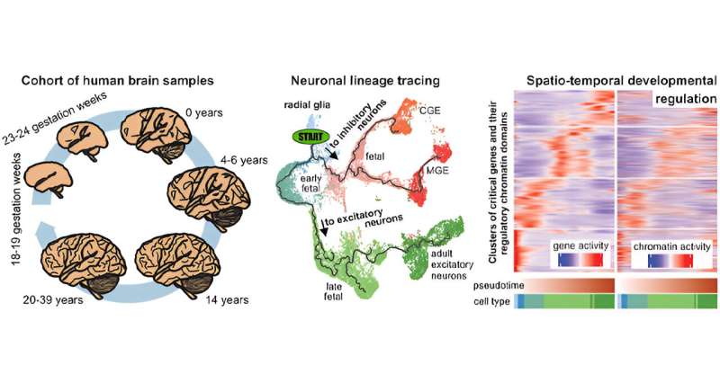
A team of researchers has created the first “multiome” atlas of brain cell development in the human cerebral cortex across six broad developmental time points from fetal development into adulthood, shedding new light on their roles during brain development and disease. “Multiome” refers to the simultaneous analysis of multiple types of genetic information within the same biological sample. They can include the genome, the DNA encoded in our cells; the transcriptome, the RNA copies that the cell makes from the genome; and the epigenome, chemical modifications and regulatory factors that determine the accessibility of chromatin.
Scientists have established how the activity of our brain during imaginary movement differs from that during real action.
A new study reveals ancestries around the world possess a shared genetic architecture for problematic alcohol use (PAU)—habitual heavy drinking, accompanied by harmful consequences. The findings, published in Nature Medicine, could help scientists understand the genetic basis of PAU, a major cause of health problems in many age groups.
Researchers have demonstrated that differences in the gut microbiome are associated with overall cognitive function and brain structure in healthy children.
In an innovative study, researchers have conducted the first systematic investigation of the effects of cognitive fatigue by using two different tasks across three distinct populations: multiple sclerosis, traumatic brain injury, and controls.
A new study details that markers of brain injury are present in the blood many months after COVID-19 infection, despite inflammation blood tests being normal.
Finally this week, new research investigates the impact of binaural beat (BB) on language skills. BB is a sound that occurs when two slightly mismatched pure tones are heard. There is a growing interest in using BB as a non-invasive neuromodulation to enhance cognitive performance.
This video delves into a groundbreaking study revealing how experienced meditators can voluntarily enter states of deep unconsciousness and reawaken with heightened mental clarity.
These states are sometimes called “cessations” or “nirvana with remainder” in Buddhist terminology. Cessation refers to the temporary suspension of the ordinary flow of consciousness. It is considered a deep state of tranquility where the usual mental activities come to a temporary halt. This is often described as a profound stillness or emptiness.
After experiencing cessation, practitioners often report a profound sense of clarity, heightened awareness, and a deep understanding of the nature of the mind and reality. This clarity is said to result from the temporary suspension of normal cognitive processes, allowing for a direct, unmediated perception of reality.
Examples of inputs and outputs from the MADRC dataset.
Researchers have developed a suite of free tools for analyzing vast amounts of brain dissection photographs at brain banks worldwide to enhance understanding of neurodegenerative diseases.
A new study reveals a strong link between regular physical activity and enhanced brain health. Analyzing MRI scans from 10,125 individuals, researchers found that exercise, even moderate exercise like walking, is associated with increased brain volumes in crucial areas like gray matter, white matter, and the hippocampus. The study underscores exercise’s role in reducing dementia risk and maintaining brain size.
Scientists have discovered that a part of the brain associated with working memory and multisensory integration may also play an important role in how the brain processes social cues.
In a first-of-its-kind study published in Nature, researchers recorded activity from hundreds of individual neurons while participants listened to spoken sentences, giving us an unprecedented view into how the brain analyzes the sounds in words.
A new study has unveiled three distinct cognitive deficits contributing to reading difficulties in individuals with left-sided neglect dyslexia, a condition that often follows a right-hemisphere stroke.
Researchers have unveiled a significant similarity between AI memory processing and human hippocampal functions. This discovery, bridging AI and neuroscience, highlights a parallel in memory consolidation – a process crucial in transforming short-term to long-term memories – in both AI models and the human brain.
A new study highlights the significant role of imagination in evoking empathy and driving prosocial behaviour.
A so-called pathological protein long associated with Parkinson’s disease has been found in a new study to trigger cells to increase protein synthesis, an event that eventually kills the subset of brain cells that die off in this neurodegenerative condition.
A new study presents a promising treatment for restoring the sense of smell in long-COVID patients.
Researchers have found that amyloid oligomers play a role in speeding up mitochondrial energetics during the early stages of Alzheimer’s, in contrast to what has been previously found in more advanced Alzheimer’s brain tissues. The results are publishedin Nature Communications.
Research led by the Karolinska Institutet, Sweden, has found an increased risk of cardiovascular disease associated with long-term ADHD medication use.
New research has uncovered a potential early marker for autism in infants: abnormally enlarged perivascular spaces (PVS) in the brain. The study found that infants with enlarged PVS had a 2.2 times greater chance of developing autism compared to those with the same genetic risk. The researchers followed infants with a higher likelihood of autism due to having an older sibling with the condition.
Signs of injury to the brain’s white matter called white matter hyperintensities, as seen on brain scans, may be tied more strongly to vascular risk factors, brain shrinkage, and other markers of dementia in former tackle football players than in those who did not play football, according to a study published in Neurology.
Artificial intelligence, coupled with data from an iPad coloring game, could assist in early diagnosis of autism, a new study shows.
A review in the Journal of Internal Medicine explores the potential of non-invasive interventions such as light, sound, and magnets to stimulate gamma brain waves for the treatment of Alzheimer’s disease. Such strategies may be beneficial because Alzheimer’s disease is characterized by reduced fast brain oscillations in the gamma range (30–100 Hz).
Finally this week, researchers have made a significant breakthrough in understanding the genetic basis of anxiety disorders (ADs), which affect over 280 million people globally.